Understanding Tetralogy of Fallot
Getting to know this congenital heart defect
Among the more serious congenital heart defects ranks Tetralogy of Fallot (TOF) which is a rare disease present at birth. It affects the normal flow of blood through the heart and is one of the most common causes of “blue baby syndrome,” where babies have a bluish tint to their skin due to lack of oxygen. This condition accounts for about 10% of all congenital heart defects.
Four Defects – a defining characteristic
In this disease four key heart abnormalities occur together giving rise to the term “tetralogy”. Let’s take a look at them:
- Ventricular Septal Defect (VSD): This refers to a hole in the wall (septum) that separates the two lower chambers (ventricles) of the heart. This allows oxygen-rich and oxygen-poor blood to mix.
- Pulmonary Stenosis: In this there is a narrowing of the pulmonary valve and artery reducing blood flow from the right ventricle to the lungs.
- Right Ventricular Hypertrophy: The right ventricle in the afflicted baby becomes thickened because it has to work harder to pump blood through the narrowed pulmonary valve.
- Overriding Aorta: The aorta is positioned directly above the ventricular septal defect, allowing oxygen-poor blood from the right ventricle to flow into it, instead of going to the lungs.
What are the causes and risk factors?
While it is difficult to pinpoint the exact cause of Tetralogy of Fallot, it is believed to result from a combination of genetic and environmental factors. In fact, it may be associated with certain genetic disorders like DiGeorge syndrome or Down syndrome. Maternal factors such as poor nutrition, diabetes or exposure to harmful substances during pregnancy may also increase the risk of this disease.
What are the symptoms?
One of the most noticeable symptoms of this disease is cyanosis – a bluish colouration of the lips, fingers and toes, especially during feeding or crying. Some of the other symptoms are:
- Difficulty in breathing
- Poor weight gain
- Fatigue or fainting during activity
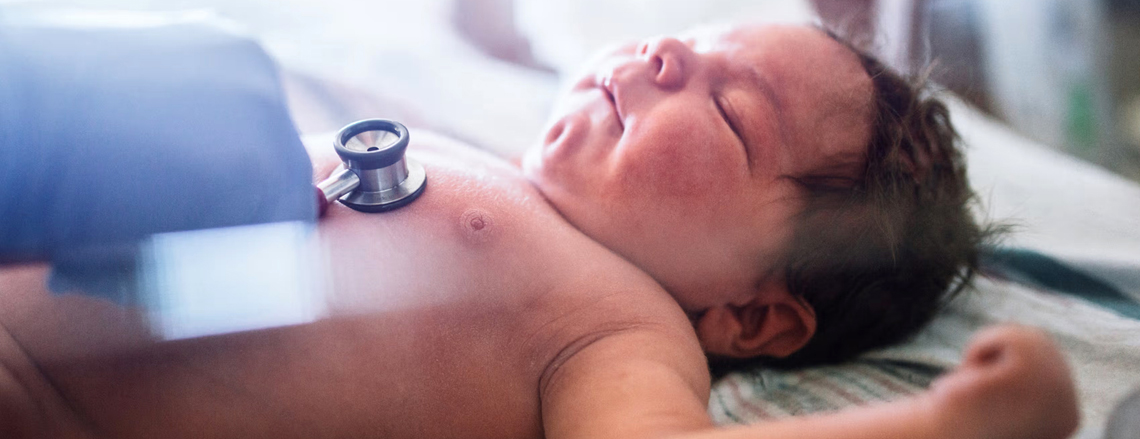
- Heart murmur (a whooshing sound that can be heard with a stethoscope)
- Occurrence of “Tet spells” which are sudden episodes of deep blue skin, nails and lips due to a drop in oxygen levels
Diagnosis protocols
This disease is not one to be taken lightly and TOF is often diagnosed in infancy or even before birth through prenatal ultrasound. After birth, doctors may use:
- Echocardiogram: To view the heart’s structure and function
- Electrocardiogram (ECG): To check the heart’s electrical activity
- Chest X-ray: To see the shape and size of the heart
- Cardiac catheterisation: For detailed imaging and pressure measurements inside the heart
What is the right course of treatment?
The course of treatment in regard to this disease is very clear with surgery being the only effective treatment. Most babies undergo open-heart surgery within the first year of life. The procedure involves closing the ventricular septal defect and widening the narrowed pulmonary valve or artery. In some cases, a temporary surgery may be done first to improve oxygen flow before the full repair.
After surgery, most children live healthy lives, though some may need further interventions later in life. Of course, regular follow-ups with a cardiologist are essential.
Beating the ailment
As mentioned, it is possible to combat the disease with the right treatment and surgery. Timely surgical treatment and ongoing medical care ensures that the prognosis for children with Tetralogy of Fallot is generally good. Many patients go on to lead active, fulfilling lives. Early diagnosis, careful monitoring and advances in paediatric cardiology have significantly improved outcomes for affected children.